Protective effects of atung (Parinarium glaberrimum) seed extract against streptozotocin-induced pancreatic damage: an in vivo experimental study

Downloads
Background: Hyperglycemia, characterized by elevated blood glucose levels, leads to oxidative stress and pancreatic β-cell dysfunction. Streptozotocin (STZ)-induced hyperglycemia in mice is widely used to model pancreatic injury and evaluate antihyperglycemic agents. Atung (Parinarium glaberrimum) seed extract contains antioxidant compounds; however, its effects on pancreatic histopathology remain unclear.
Objective: This study aimed to investigate the effect of atung seed extract on pancreatic histopathology in STZ-induced hyperglycemic mice, focusing on islet diameter and histopathological damage.
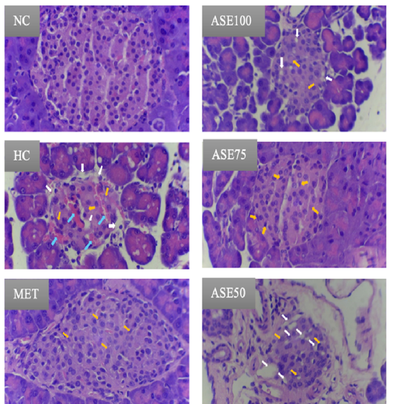
Methods: Twenty-four male Mus musculus (BALB/c) mice were randomly allocated into six groups: normal control, hyperglycemic control, metformin-treated, and three atung seed extract groups (100%, 75%, and 50%). Hyperglycemia was induced using intraperitoneal streptozotocin (40 mg/kgBW). Treatments were administered orally for 21 days. Blood glucose levels were reassessed on day 22 prior to euthanasia. Pancreatic tissues were collected, fixed in 10% neutral buffered formalin, and stained with hematoxylin–eosin.
Results: Islet diameter differed significantly among groups (p < 0.05). The ASE75 group showed a mean diameter of 133.01 µm and a median damage score of 1 (IQR 0), comparable to the metformin group (146.61 µm; 0.5 [IQR 1]), whereas the hyperglycemic control group showed severe atrophy (89.02 µm; 3 [IQR 0]).
Conclusion: Atung seed extract at a 75% concentration effectively preserved pancreatic β-cell structure, as indicated by increased islet diameter and reduced histopathological damage, supporting its potential as a natural antioxidant-based adjuvant therapy for hyperglycemia management.

This work is licensed under a Creative Commons Attribution-NonCommercial 4.0 International License.
Authors who publish with this journal agree to the following terms:Authors retain copyright and grant the journal right of first publication with the work simultaneously licensed under a Creative Commons Attribution-NonCommercial 4.0 International License that allows others to share the work with an acknowledgement of the work's authorship and initial publication in this journal.
Authors are able to enter into separate, additional contractual arrangements for the non-exclusive distribution of the journal's published version of the work (e.g., post it to an institutional repository or publish it in a book), with an acknowledgement of its initial publication in this journal.
Authors are permitted and encouraged to post their work online (e.g., in institutional repositories or on their website) prior to and during the submission process, as it can lead to productive exchanges, as well as earlier and greater citation of published work (See The Effect of Open Access).
This work is licensed under a Creative Commons Attribution-NonCommercial 4.0 International License.