Computed tomography scan imaging in case of destroyed lung post-tuberculosis

Downloads
Background: Destroyed lung is a radiological term describing severe and irreversible damage to the lung parenchyma, most commonly resulting from TB. This condition poses significant clinical challenges and requires advanced imaging for timely diagnosis and appropriate management.
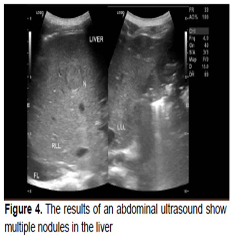
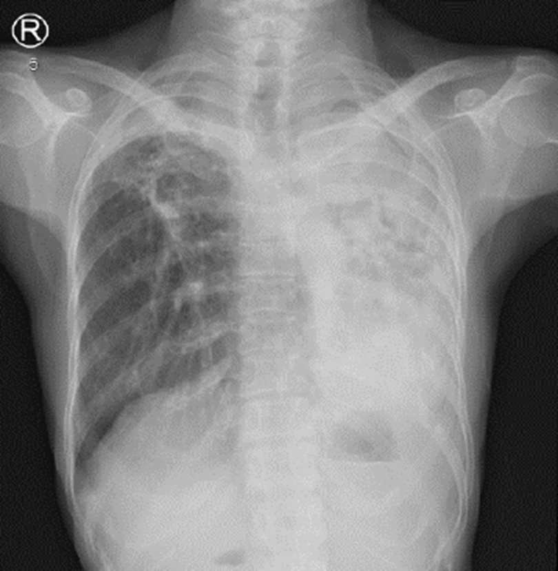
Case Presentation: This report presents three cases illustrating the clinical and radiological features of destroyed lung secondary to tuberculosis. The first case involves a 45-year-old woman with a prior history of TB who presented with facial swelling, dyspnea, and chronic cough; imaging revealed extensive cavitary lesions and destruction of the left lung. The second case describes a 25-year-old man with active TB who experienced worsening shortness of breath and night sweats; CT imaging showed a left fluidopneumothorax, multiple bullae, and bilateral cavitary lesions, indicating destroyed lung with secondary infection. The third case features a 46-year-old woman with chronic respiratory symptoms and right-sided pleural effusion; imaging demonstrated right lung destruction, bilateral fibrotic changes, and pulmonary artery hypertension.
Conclusion: Destroyed lung remains a significant post-tuberculosis complication requiring prompt diagnosis and multidisciplinary management. Radiologists play a critical role in identifying characteristic imaging findings and reviewing prior imaging to support clinicians in developing appropriate treatment strategies.
1. Osarenkhoe J, Aiwuyo H, Aisosa O, Ejiroghene U. Destroyed Lung Syndrome: A Review of 31 Published Cases. Open Journal of Respiratory Diseases. 2022;12(02):37-43. doi:10.4236/ojrd.2022.122003
2. Jeong YJ, Lee KS. Pulmonary Tuberculosis: Up-to-Date Imaging and Management. American Journal of Roentgenology. 2008;191(3):834-844. doi:10.2214/AJR.07.3896
3. Isbaniah F, Burhan E, Sinaga BYM, et al. Tuberkulosis Pedoman Diagnosis Dan Penatalaksaan Di Indonesia. 2 Revision. Jakarta: Perhimpunan Dokter Paru Indonesia; 2021.
4. Carette MF, Blanchon F, Milleron B, Brocard H. [Destroyed lung (author’s transl)]. Sem Hop. 55(17-18):843-853.
5. Mısırlıoğlu AK. Factors affecting complication rates of pneumonectomy in destroyed lung. The Turkish Journal of Thoracic and Cardiovascular Surgery. 2018;26(2):272-278. doi:10.5606/tgkdc.dergisi.2018.14635
6. Han D, Lee HY, Kim K, Kim T, Oh YM, Rhee CK. Burden and clinical characteristics of high grade tuberculosis destroyed lung: a nationwide study. J Thorac Dis. 2019;11(10):4224-4233. doi:10.21037/jtd.2019.09.63
7. Patil S, Narkar S, Raka V, Dahiphale J, Choudhari S, Gondhali G. ‘Destroyed lung’ as Post Tuberculosis Sequel: A Preventable Stigma of ‘disease of concern’ of Millennium! Saudi Journal of Medicine. 2023;8(03):112-119. doi:10.36348/sjm.2023.v08i03.007
8. Ravimohan S, Kornfeld H, Weissman D, Bisson GP. Tuberculosis and lung damage: from epidemiology to pathophysiology. European Respiratory Review. 2018;27(147):170077. doi:10.1183/16000617.0077-2017
9. Rhee CK, Yoo KH, Lee JH, et al. Clinical characteristics of patients with tuberculosis-destroyed lung. The International Journal of Tuberculosis and Lung Disease. 2013;17(1):67-75. doi:10.5588/ijtld.12.0351
10. Yadav S. Destroyed Lung Syndrome in a Young Indian Male: A Case Report. Cureus. April 2023. doi:10.7759/cureus.38174
11. Radzikowska E. Pulmonary Langerhans’ Cell Histiocytosis in Adults. Adv Respir Med. 2017;85(5):277-289. doi:10.5603/ARM.a2017.0046
12. Varona Porres D, Persiva O, Pallisa E, Andreu J. Radiological findings of unilateral tuberculous lung destruction. Insights Imaging. 2017;8(2):271-277. doi:10.1007/s13244-017-0547-4
13. Ruan H, Liu F, Han M, Gong C. Incidence and risk factors of postoperative complications in patients with tuberculosis-destroyed lung. BMC Pulm Med. 2021;21(1):273. doi:10.1186/s12890-021-01641-0
14. Jullien S, Jain S, Ryan H, Ahuja V. Six-month therapy for abdominal tuberculosis. Cochrane Database of Systematic Reviews. 2016;2016(11). doi:10.1002/14651858.CD012163.pub2
15. Aurangabadkar GM, Choudhary SS, Khan SM, Jadhav US, Wagh PB, Ali SN. Post-tubercular Unilateral Lung Destruction: A Complicated Case. Cureus. June 2023. doi:10.7759/cureus.40035
16. Mehta K, Lekshmi R. Navigating Complex Pulmonary Hypertension: A Case of Post-tubercular Lung Disease and Atrial Septal Defect. JOURNAL OF CLINICAL AND DIAGNOSTIC RESEARCH. 2024. doi:10.7860/JCDR/2024/68053.19239
17. Olguntürk R, Tunaoğlu FS, Gökgöz L, Memiş L, Kula S. Congenital heart disease and pulmonary tuberculosis. Open Medicine. 2010;5(2):172-175. doi:10.2478/s11536-009-0061-2
18. Bansal N, Arunachala S, Kaleem Ullah M, et al. Unveiling Silent Consequences: Impact of Pulmonary Tuberculosis on Lung Health and Functional Wellbeing after Treatment. J Clin Med. 2024;13(14):4115. doi:10.3390/jcm13144115
19. Allwood BW, Byrne A, Meghji J, Rachow A, van der Zalm MM, Schoch OD. Post-Tuberculosis Lung Disease: Clinical Review of an Under-Recognised Global Challenge. Respiration. 2021;100(8):751-763. doi:10.1159/000512531
20. Cupido G, Günther G. Post tuberculosis lung disease and tuberculosis sequelae: A narrative review. Indian Journal of Tuberculosis. 2024;71(1):64-72. doi:10.1016/j.ijtb.2023.04.001
21. Bhende V V, Chaudhary A, Madhusudan S, et al. A Global Bibliometric Analysis of the Top 100 Most Cited Articles on Early Thoracotomy and Decortication in Pleural Empyema. Cureus. October 2024. doi:10.7759/cureus.72800
22. Dantis K, Kumar Dewan R. Surgical outcomes and the factors affecting lung expansion following open window thoracostomy in chronic tuberculous empyema with destroyed lung. Asian Cardiovasc Thorac Ann. 2022;30(6):696-705. doi:10.1177/02184923221104431
23. Chu H, Li B, Zhao L, et al. Tree-in-bud pattern of chest CT images for diagnosis of Mycobacterium abscesses. Int J Clin Exp Med. 2015;8(10):18705-18712.
24. Im JG, Itoh H. Tree-in-Bud Pattern of Pulmonary Tuberculosis on Thin-Section CT: Pathological Implications. Korean J Radiol. 2018;19(5):859. doi:10.3348/kjr.2018.19.5.859
25. Liu L, Wang X, Luo L, Liu X, Chen J. Risk Factors of Tuberculosis Destroyed Lung in Patients with Pulmonary Tuberculosis and Structural Lung Diseases: A Retrospective Observational Study. Risk Manag Healthc Policy. 2024;Volume 17:753-762. doi:10.2147/RMHP.S448765
26. Gai X, Allwood B, Sun Y. Post-tuberculosis lung disease and chronic obstructive pulmonary disease. Chin Med J (Engl). 2023;136(16):1923-1928. doi:10.1097/CM9.0000000000002771
27. Yoon S, Mihn DC, Song JH, Kim SA, Yim JJ. Evolution of Interferon-Gamma Release Assay Results and Submillisievert Chest CT Findings among Close Contacts of Active Pulmonary Tuberculosis Patients. Tuberc Respir Dis (Seoul). 2020;83(4):283-288. doi:10.4046/trd.2020.0038
28. Urbanowski ME, Ordonez AA, Ruiz-Bedoya CA, Jain SK, Bishai WR. Cavitary tuberculosis: the gateway of disease transmission. Lancet Infect Dis. 2020;20(6):e117-e128. doi:10.1016/S1473-3099(20)30148-1
29. Ko JM, Kim KJ, Park SH, Park HJ. Bronchiectasis in active tuberculosis. Acta radiol. 2013;54(4):412-417. doi:10.1177/0284185113475796
30. Moule MG, Cirillo JD. Mycobacterium tuberculosis Dissemination Plays a Critical Role in Pathogenesis. Front Cell Infect Microbiol. 2020;10. doi:10.3389/fcimb.2020.00065
31. Nachiappan AC, Rahbar K, Shi X, et al. Pulmonary Tuberculosis: Role of Radiology in Diagnosis and Management. RadioGraphics. 2017;37(1):52-72. doi:10.1148/rg.2017160032
Copyright (c) 2025 MEDISAINS

This work is licensed under a Creative Commons Attribution-NonCommercial 4.0 International License.
Authors who publish with this journal agree to the following terms:Authors retain copyright and grant the journal right of first publication with the work simultaneously licensed under a Creative Commons Attribution-NonCommercial 4.0 International License that allows others to share the work with an acknowledgement of the work's authorship and initial publication in this journal.
Authors are able to enter into separate, additional contractual arrangements for the non-exclusive distribution of the journal's published version of the work (e.g., post it to an institutional repository or publish it in a book), with an acknowledgement of its initial publication in this journal.
Authors are permitted and encouraged to post their work online (e.g., in institutional repositories or on their website) prior to and during the submission process, as it can lead to productive exchanges, as well as earlier and greater citation of published work (See The Effect of Open Access).
This work is licensed under a Creative Commons Attribution-NonCommercial 4.0 International License.